
Figure 2.
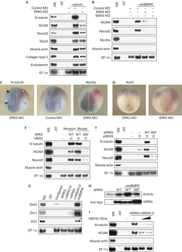
Involvement of the MEK5–ERK5 pathway in neural differentiation. (A) Control morpholino oligonucleotide (MO) or ERK5 MO (20 ng in total) was injected into each blastomere of four-cell-stage embryos. Animal caps were treated with 10 ng/ml activin and cultured to stage 20. The expression of marker genes was examined. Elongation factor-1α (EF-1α) served as a loading control; WE, whole embryo. (B) Dominant-negative bone morphogenetic protein receptor (dnBMPR) messenger RNA (0.4 ng) with control MO (160 ng), ERK5 MO (20 ng) or ERK5 MO (160 ng) was injected and animal caps from the embryos were analysed by reverse transcription–PCR (RT–PCR). (C,D) ERK5 MO or control MO (20 ng) was injected into the right blastomeres of four-cell-stage embryos. The expression pattern of N-tubulin or MyoDa (neurula stages) (C) or SoxD (gastrula stage) (D) was analysed by whole-mount in situ hybridization. Nuclear localization signal–β-galactosidase mRNA was co-injected and used as a tracer (red staining). Dorsal views of embryos are shown with the anterior side at the top. (E,F) Indicated sets of mRNAs (1.0 ng) were injected and animal caps from the embryos were analysed by RT–PCR; WT, wild type. (G) Indicated sets of mRNAs were injected. Animal caps were cultured to stage 12 and analysed by RT–PCR. (H) Myc–xERK5 (Xenopus orthologue of ERK5) or Myc–xERK5 AEF mRNA (1.0 ng) was injected with dnBMPR mRNA (0.4 ng) and animal caps were cultured to stage 14. Activity of xERK5 was measured by kinase assays. The amounts of immunoprecipitated xERK5 were detected by anti-Myc antibody. (I) xERK5 and xMEK5 D (constitutively active xMEK5) mRNAs were injected with MEF2C R24L mRNA (1.0 or 2.0 ng) and animal caps were subjected to RT–PCR.