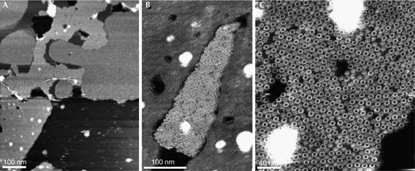
Figure 3.

Overview atomic force microscopy topographs of reconstituted c-subunit oligomers of F1F0 ATP synthase from Spirulina platensis. (A) Densely distributed membrane patches adsorbed onto mica showing empty lipid membranes and densely packed c oligomers (bright corrugated region). A higher magnification shows a densely packed assembly of oligomeric rings (centre of images B,C). Full grey level ranges correspond to vertical dimensions of 15 nm (A) and 5 nm (B,C).
