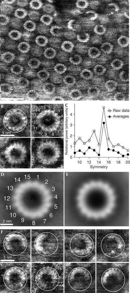
Figure 4.

High-resolution atomic force microscopy topographs of reconstituted rotor rings. (A) Survey of an almost two-dimensional crystalline arrangement of c rings in the membrane. (B) At a higher magnification, the c subunits of single rotors could be directly observed. The c rings were selected from different topographs. (C) Rotational power spectra calculated from single rings with sufficiently high signal-to-noise ratio (such as those shown in B) and from the three major averages (as shown in D) found by reference-free averaging, show a clear symmetry. (D) Average, generated by reference-free translational and rotational alignment of 90 complete c rings. (E) A 15-fold symmetrized topography of average shown in (C). (F) Gallery of incomplete rotors showing the same diameter (white circles) as the complete rotors (B). Full grey level ranges correspond to vertical scales of 4 nm (A) and 3.5 nm (B,D–F).
