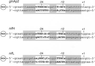
Figure 1.
Three different ROX-labeled DNA duplexes were used in the binding studies, the glnAp2 promoter sequence from E.coli and the nifH and nifL promoters from K.pneumoniae. The nucleotides that fit the –24/–12 consensus sequence for RNAP·σ54-specific promoters are in bold (1). The RNAP binding region from about –34 close to the transcription start site at position –2 is shaded in gray and has been derived from footprinting studies (44,45). Positions +1, –12 and –24 relative to the RNA transcript start site are indicated.

