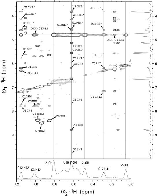
Figure 2.
NOESY spectra of the 3′SL from human U4 snRNA in H2O buffer at 20°C acquired with a 250 ms mixing time. The bottom trace shows a slice through the water frequency with the exchange peaks labeled. The trace on the right shows a slice through the frequency from the 2′-OH of U10. The vertical lines are drawn through the frequencies of C12 H41 and H42 and the frequencies of three hydroxyl protons. Two broad, unlabeled exchange peaks in the bottom panel come from the unassigned amino groups of adenine or guanine. The NOE cross-peaks from U10 2′-OH establish the assignment and establish that the UACG tetraloop in 3′SL is stabilized by a hydrogen bond to O6 of G13. A similar hydrogen bond has been observed previously in the NMR structure of UUCG tetraloops. The pattern of hydrogen bonds that stabilize the UACG tetraloop is shown in Figure 4A.

