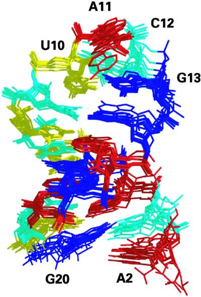
Figure 3.

Overlay of the 10 structures of lowest total energy determined from the refinement protocol based on 200 distance restraints derived from NMR spectroscopic data. The stem region adopts an A-form helix while the UACG tetraloop is stabilized by a network of hydrogen bonds as well as base stacking. Adenines are colored in red, guanines in blue, uracils in yellow and cytosines in cyan. Coordinates of the 10 structures, distance restraints, integrated NOE intensities and proton chemical shifts have been deposited in the Protein Data Bank (accession number 1MFJ).
