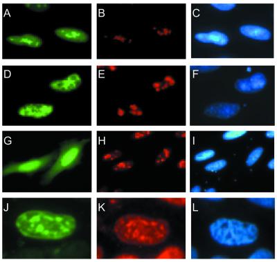
Figure 3.
Nuclear localisation of the hFPG1–EGFP and hFPG2–EGFP fusion proteins. Exponentially growing asynchronous HeLa S3 cells were transiently transfected with constructs expressing hFPG1–EGFP (A–C), EGFP–hFPG2 (D–F and J–L) and pEGFP–N1 (G–I). The cells were imaged directly by fluorescence microscopy for EGFP detection (green, A, D, G and J). Nucleolin (red, B, E and H) and RPA (red, K) were both visualised by immunofluorescence with specific monoclonal antibodies followed by Alexa-conjugated secondary antibodies. DNA was stained by DAPI (blue, C, F, I and L).

