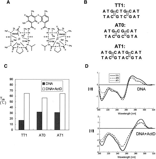
Figure 1.
(A) Chemical structure of ActD. The cyclic pentapeptide attached to the quinonoid ring and the benzenoid ring of the phenoxazone group are labeled α and β, respectively. (B) DNA duplexes that were used in this study include AT0, AT1 and TT1 (rectangles represent the binding sites for the phenoxazone ring of ActD). (C) The effect of ActD on the Tm values of AT0, AT1 and TT1 (4 µM) in standard buffer. (D) CD spectra of AT0, AT1 and TT1 (4 µM) in standard buffer alone (top) and with 10 µM ActD are graphed (bottom). The CD spectra of ActD–DNA complexes were obtained by subtracting the spectrum of ActD.

