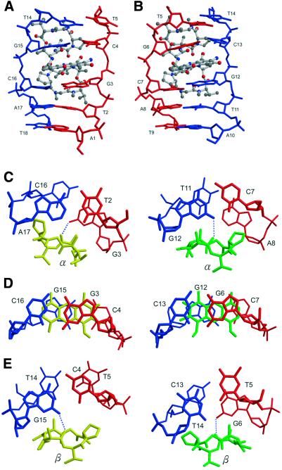
Figure 4.
Close-up side view of the ActD–TGCAT part (ActD in ball-and-stick and DNA in skeletal representation). The two phenoxazone rings are intercalated individually into the (G3pC4)·(G15pG16) step in (A) and the (G6pC7)·(G12pC13) step in (B), respectively. (C–E) The detailed conformation showing the stacking interactions in the ActD–DNA complex at various base pair steps of the refined structure. The hydrogen bonding between the oxygen atom of the threonine carbonyl group and the hydrogen atom of the guanine N2 amino group is marked by blue dotted lines.

