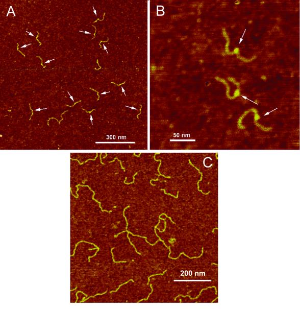
Figure 2.
AFM images of the 447 bp fragment containing a hemiknot structure. (A and B) Large-scale image and a zoomed image with three clearly seen blobs, respectively. The blobs on the filaments are indicated with arrows. (C) AFM images of a control sample, an untreated 447 bp fragment. The samples were deposited onto APS-mica and imaged in air with TM AFM after the rinse–dry step.

