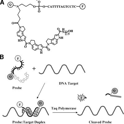
Figure 3.
(A) The structure of a fluorogenic MGB-probe conjugate. Q is a diazine analog, F is fluorophore. (B) Illustration of the hybridization of the MGB-Q-ODN-F conjugate to its target. The MGB binds in the minor groove to stabilize the duplex. Taq polymerase cleaves the 3′-MGB-Q-ODN-F conjugate during PCR to generate a fluorescent signal.

