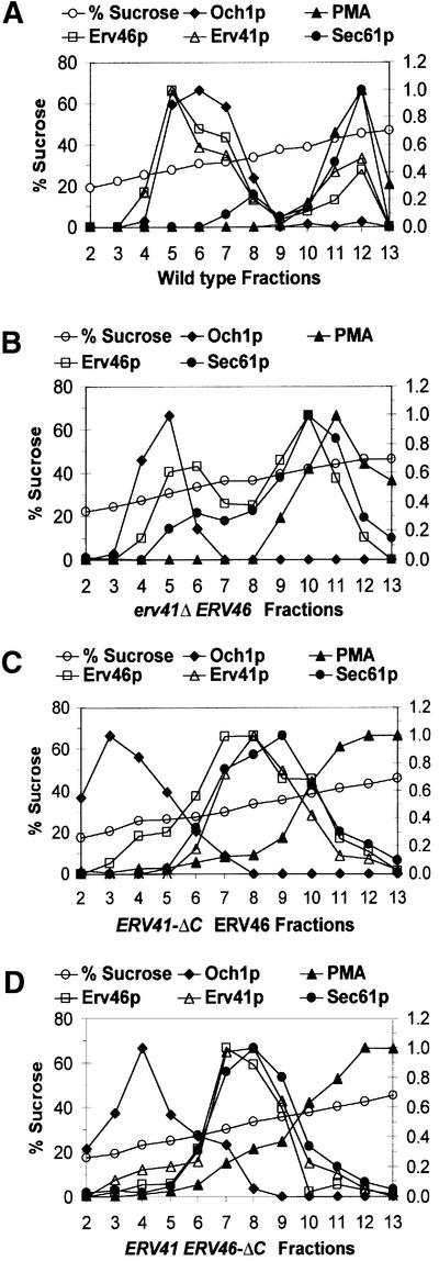
Fig. 3. Subcellular localization of Erv41p and Erv46p depends on the presence of both C-terminal tail sequences. Cell lysates were resolved on 18–60% (w/v) sucrose gradients and fractions were collected from the top. Relative amounts of the ER marker Sec61p, the Golgi marker Och1p, the plasma membrane marker PMA1, Erv41p and Erv46p were quantified by densitometry of immunoblots. Sucrose percentages were determined by refractometry. In the first experiment, (A) wild-type (FY834) and (B) erv41Δ (CBY797) strains were compared. In the second experiment, (C) ERV41-ΔC (CBY1199) and (D) ERV46-ΔC (CBY1200) were analyzed.

An official website of the United States government
Here's how you know
Official websites use .gov
A
.gov website belongs to an official
government organization in the United States.
Secure .gov websites use HTTPS
A lock (
) or https:// means you've safely
connected to the .gov website. Share sensitive
information only on official, secure websites.
