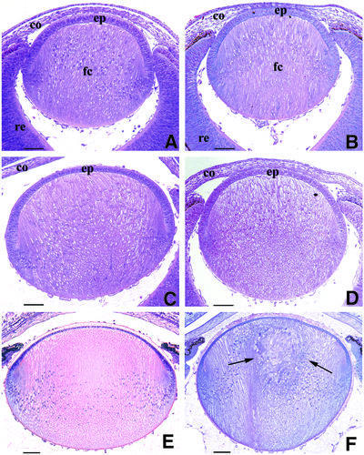
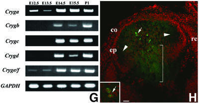
Fig. 1. Histochemical analysis of the appearance of the cataract phenotype in the Crygbnop mouse compared with the wild-type from E14.5 to birth (P1). At E14.5, the primary fibre cells fill the lumen of the lens vesicle and the first secondary fibre cells are formed at the lens cortex in both the wild-type (A) and the Crygb (B) lenses. The γBnop-crystallin is first expressed at this developmental time point [see (G) and (H) below]. (C and D) At E15.5, the first phenotypic changes are obvious. The fibre cells appear swollen and the lens is clearly smaller (D) as compared with the wild-type lens (C). This process continues in the newborn mouse where, in contrast to the wild-type lens (E), the centre of the Crygbnop lens remains opaque and the primary lens fibre cells become completely disorganized (F, arrows). (G) All six Cryg cDNAs were amplified from E12.5 to P1 from mouse tissues (E12.5–E14.5, head; E15.5, whole eyes; P1, lens). The individual transcripts are indicated along the vertical and the developmental times along the horizontal. The expression of GAPDH was used as a positive control. Cryge/f and Cryga are expressed by E12.5, whereas Crygb is first detected at E14.5. (H) Polyclonal antibodies specific to γBnop-crystallin (Klopp et al., 1998) were used to detect the mutant protein in E14.5 lenses. E13.5 lenses were negative (data not shown), but in the E14.5 lens, γBnop-crystallin (green channel) is present both as nuclear inclusions (arrow and inset) and in the lens fibre cell cytoplasm (bracket) where it is not present as inclusions. This section has been counterstained with propidium iodide (red channel) to locate the cell nuclei. ep, lens epithelium; fc, fibre cell; re, retina; co, cornea. Scale bars = 250 µm in (A–F) and 30 µm in (H).

An official website of the United States government
Here's how you know
Official websites use .gov
A
.gov website belongs to an official
government organization in the United States.
Secure .gov websites use HTTPS
A lock (
) or https:// means you've safely
connected to the .gov website. Share sensitive
information only on official, secure websites.

