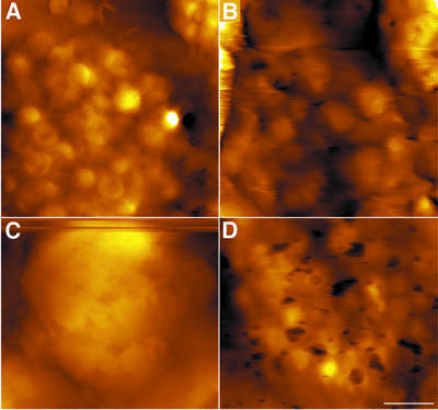
Fig. 4. Grana unstacking imaged on native, hydrated thylakoids. An increase in grana size is observed. Native thylakoid membranes (A) were subjected to 50 (B) or 100 (C) mM EDTA. For re-stacking, samples treated with 100 mM EDTA were incubated in a medium containing 5 mM Mg2+ (D). Evidently, the original membrane organization has been restored. Because repetitive scans over long periods damaged the thylakoids, images were obtained from different samples. The scale bar is 1 µm.

An official website of the United States government
Here's how you know
Official websites use .gov
A
.gov website belongs to an official
government organization in the United States.
Secure .gov websites use HTTPS
A lock (
) or https:// means you've safely
connected to the .gov website. Share sensitive
information only on official, secure websites.
