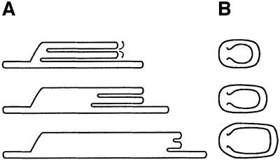
Fig. 6. A model of membrane unstacking. (A) Cross-section of the thylakoid stacks. (B) Top view. Unstacking begins by evagination of the membranes located at the stromal-exposed edges of the appressed grana domains (top). As unfolding proceeds, membranes are pulled from the stack’s interior towards the edges, allowing for an increase in lateral dimensions with no change in height (middle and bottom). Eventually, the stacks merge completely with the neighbouring stroma lamellae, and grana topology is lost (Figure 4C).

An official website of the United States government
Here's how you know
Official websites use .gov
A
.gov website belongs to an official
government organization in the United States.
Secure .gov websites use HTTPS
A lock (
) or https:// means you've safely
connected to the .gov website. Share sensitive
information only on official, secure websites.
