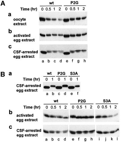
Fig. 4. Degradation and phosphorylation of E.coli-derived Xenopus MOS and its derivatives in extracts from Xenopus oocytes, activated eggs and CSF-arrested eggs. (A) Pro-Ser-MOSfhk and Gly-Ser- MOSfhkP2G in oocyte extract (a), activated egg extract (b) and CSF-arrested egg extract (c). Degradation (monitored through immunoblotting) and phosphorylation (detected through shifts in electrophoretic mobility) as a function of time after the addition of MOS to an extract. (B) Pro-Ser-MOSfhk, Gly-Ser-MOSfhkP2G and Pro-Ala-MOSfhkS3A in activated egg extract and CSF-arrested egg extract. In these experiments, a MOS test protein was pre-incubated, at 100 µg/ml, in 30 µl of the CSF-arrested egg extract for 1 h (a), followed by the addition of a resulting sample to either the activated egg extract (b) or CSF-arrested egg extract (c), to a final total volume of 25 µl and a final MOS concentration of ∼25 µg/ml.

An official website of the United States government
Here's how you know
Official websites use .gov
A
.gov website belongs to an official
government organization in the United States.
Secure .gov websites use HTTPS
A lock (
) or https:// means you've safely
connected to the .gov website. Share sensitive
information only on official, secure websites.
