Figure 1.

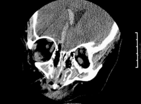
Coronal CT; blood and air extending in a straight line from the cribriform plate to the right lateral ventricle, with blood filling the right lateral ventricle. (Note: blood in the third ventricle was also seen but not shown here.)

Coronal CT; blood and air extending in a straight line from the cribriform plate to the right lateral ventricle, with blood filling the right lateral ventricle. (Note: blood in the third ventricle was also seen but not shown here.)