
Figure 1.
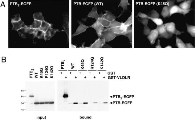
Membrane localization and VLDLR binding of the PTB/PI domain of Dab1. C-terminal EGFP fusions of wildtype or various mutant forms of the Dab1 PTB domain were expressed in 293T cells. (A) Epifluorescence images. Wildtype Dab1 PTB is enriched at cell edges and cell-cell junctions, indicating membrane association. K45Q mutant Dab1 PTB is not membrane associated, and a double PTB protein shows increased association. (B) Binding to the VLDLR C terminus, fused to GST, was assayed in vitro. The double PTB protein bound with higher avidity than the single PTB, and K45Q mutation had no effect on binding, in contrast to R124Q and K142Q.