Figure 6.
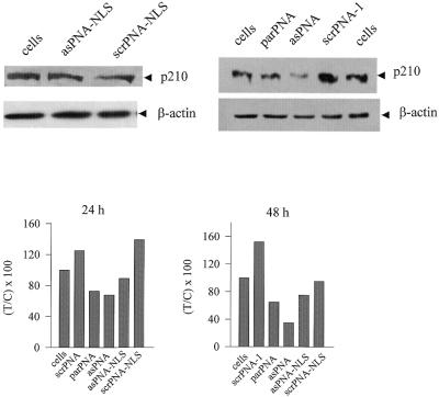
Immunoblot of KYO-1 cell lysates using anti-ABL and anti-β-actin monoclonal antibodies. KYO-1 cells were incubated with 10 µM anti sense or control PNAs and the amount of protein p210BCR/ABL was evaluated by western blot analysis, after the cells were exposed to the PNAs for 24 and 48 h. The level of β-actin in the PNA-treated cells was also measured. A typical western blot analysis of cell lysates at 48 h after PNA treatment is shown in the figure. The levels of p210BCR/ABL in lysates obtained from KYO-1 cells treated with antisense and control PNAs at 24 and 48 h are shown in the enclosed histograms. The ordinate reports the residual BCR/ABL protein expressed as percent T/C, where T is the p210BCR/ABL/β-actin ratio of PNA-treated cells and C is the p210BCR/ABL/β-actin ratio of PNA-untreated cells. The uncertainty on each value is at most 20%.

