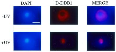
Figure 4.
Subcellular localization of D-DDB1 in Drosophila Kc cells after UV-irradiation (×1600). Bar indicates 5 µm. Kc cells were stained with DAPI (left) and antibodies against D-DDB1 (middle). Merged images are shown on the right. (Upper panels) Without UV irradiation; (lower panels) with incubation for 12 h after 70 J/m2 UV-irradiation. Note the nuclear localization of D-DDB1 after UV-irradiation.

