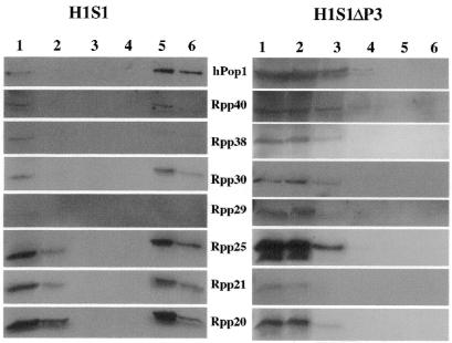
Figure 4.
Western blot analysis (Materials and Methods) of fractions from the purification step of streptavidin agarose. The blots have been probed with antibodies to the various proteins listed between the columns of figures. The left part is from HeLa cells transfected with pΔEGFP–H1S1 whereas the right part is from cells transfected with pΔEGFP–H1S1ΔP3. Lane 1, S16 extract. Lane 2, supernatant after rotation with streptavidin agarose overnight. Lane 3, flow through washed with lysis buffer. Lane 4, the eluate (0.5 mM biotin). Lane 5, the eluate (5 mM biotin). Lane 6, agarose beads after eluates.

