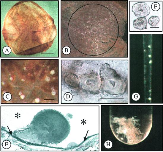
Fig. 1.
Isolation of drusen. (A) Bruch's membrane/choroid dissected from an 82-year-old donor eye. (B) Macular region of Bruch's membrane containing drusen (white particulate material), diameter = 3 mm. (C) Drusen on surface of Bruch's membrane. (D) Drusen on Bruch's membrane. (E) Histological section of drusen on Bruch's membrane (embedded in plastic, stained with toluidine blue). Asterisks represent the location of the RPE before removal and the arrows indicate Bruch's membrane. (F) Isolated drusen. (G) Isolated drusen in pipette. (H) Isolated drusen in test tube. (Bars = A, 5 mm; C, 100 μm; D, 50 μm; E and F, 25 μm.)

