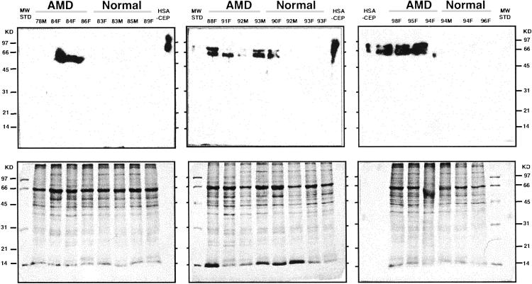
Fig. 3.
Western analysis of AMD and normal Bruch's membrane/RPE/choroid tissues. Human Bruch's membrane/RPE/choroid from the macular region of AMD and normal donor eyes was subjected to SDS/PAGE (≈20 μg protein/lane), electroblotted to poly(vinylidene difluoride), and probed with the rabbit polyclonal anti-CEP antibody to CEP adducts from docosahexaenoic acid. Human serum albumin modified with CEP (HSA-CEP, 20 ng) was used as a positive control. The age and sex of the donor eyes are listed at the top of each lane. More immunoreactivity can be seen in the AMD samples than in the normals.

