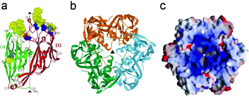
Fig. 2.
(a) Structure of the Vp54 monomer with strategic amino acids labeled. The carbohydrate moieties (yellow) and glycosylated Asn and Ser residues (blue and gray, respectively) are shown as space-filling atoms. (b) The Vp54 trimer viewed from the inside of the virus, with each monomer in a different color. (c) Surface of the Vp54 trimer also viewed from the inside of the virus, colored according to charge distribution (positive is blue, negative is red).

