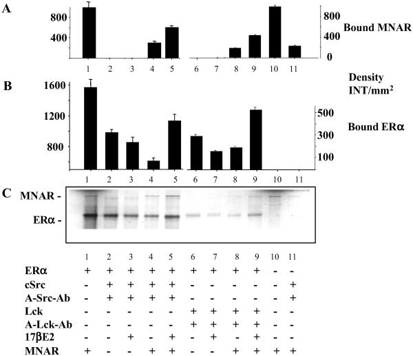
Fig 4.
ER–MNAR–Src interaction analysis. In vitro-transcribed/translated ERα with (lanes 4, 5, 8, and 9) or without (lanes 2, 3, 6, and 7) MNAR, in the presence (lanes 3, 5, 7, and 9) or absence (lanes 2, 4, 6, and 8) of 1 μM E2, were incubated with purified c-Src (0.2 μg of protein, lanes 2–5) or Lck (0.35 μg, lanes 6–9) (both from Upstate Biotechnology). Lane 1, 10% of ERα and MNAR input proteins; lane 10, 10% of MNAR input protein. Formed complexes were isolate by pull-down with anti-c-Src or anti-Lck antibodies and protein A agarose, washed, boiled in 2× SDS buffer, and resolved on an SDS/PAGE gel. Neither Src or Lck antibody nor protein A-Sepharose precipitated ERα or MNAR in the absence of Src or Lck. Band density of the gel presented in C was evaluated by using Bio-Rad software, QUANTIFY, and is plotted ± SD in A (for ERα binding) and B (for MNAR binding).

