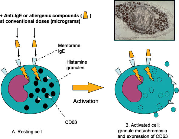
Figure 1.
Normal activation of basophil degranulation caused by anti IgE antibodies. This activation is not only driven by specific allergens, but also by the binding of antibodies against IgE heavy chains (anti-IgE) and involves changes in membrane ion fluxes (particularly calcium ions), changes in cell membrane electrical polarity, and other mechanisms that eventually lead to exocytosis and the release of mediators. Cell activation is evaluated by optical microscopy as a loss of staining properties. To be precise, loss of staining properties is not exactly the same biological phenomenon as cell degranulation, but indicates a change of the granule membrane permeability. Another typical response to activation is the increased expression of CD63 proteins, which are translocated from internal pools to the cell surface. Insert: electron microscopy of a mast cell.

