Fig 3.
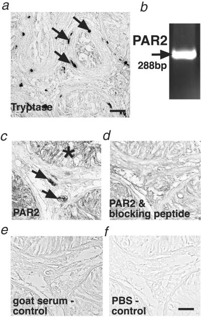
Identification of tryptase-positive mast cells and PAR2 in human testes with fibrotic changes. (a) Immunohistochemical staining of tryptase-containing mast cells in the interstitium and the wall of the seminiferous tubules (arrows) in a human testicular biopsy from a patient showing subnormal spermatogenesis and tubular fibrosis. For complete and detailed quantitative evaluation of the observed changes, see ref. 18. (Bar, 60 μm.) (b) RT-PCR analysis showing PAR2 mRNA expression in human testes. (c–f) Immunohistochemical staining showing PAR2 expression. PAR2 in a human testicular biopsy from a patient with SCO syndrome in interstitial cells (arrow) and the germinal epithelium (asterisk) (c). In a consecutive section, the PAR2 antibody was incubated previously with a specific blocking peptide to show specificity (d). For control purposes, the PAR2 antiserum was replaced by normal goat serum (e) and PBS buffer (f). (Bar, 30 μm.) Note that PAR2 RNA expression and specific immunoreaction were detected in all biopsies examined (normal group, n = 7; SCO syndrome, n = 7; GA syndrome, n = 9; and MA syndrome, n = 6).

