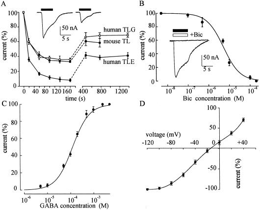
Fig 1.
Properties of GABA currents. (A) Decay of the peak GABA-current amplitude during repetitive applications of GABA (1 mM), in oocytes injected with: TLE mRNA (9 oocytes/2 frogs, 9/2; 2 patients); mouse TL mRNA (6/2; 10 mice); and TLG mRNA (6/2; 2 patients). The amplitudes were normalized to those elicited by the first applications of GABA (current range: 40–300 nA). In this and subsequent figures, the points show means ± SEM. (Inset) Sample traces of GABA currents elicited by the first (Left) and second (Right) GABA applications (filled bars) in an oocyte injected with TLE mRNA. (B) Inhibition of GABA (100 μM) currents by Bic in TLE mRNA-injected oocytes (6/2; 2 patients). The GABA currents were normalized to the currents evoked by GABA alone (Imax = −60 nA; range: 16–102 nA). (Inset) Current evoked by 1 mM GABA (filled bar), and blocked by 0.2 mM Bic (open bar). (C) GABA dose/current response relationship from TLE mRNA-injected oocytes (8/3; 3 patients). Imax = −179 nA; range: 40–330 nA. (D) GABA current/voltage relationship in TLE-oocytes (6/2; 2 patients). The GABA (100 μM) currents were normalized to those evoked at −120 mV (−115 nA; range: 70–210 nA). Reversal potential (Vrev) = −14.5 mV.

