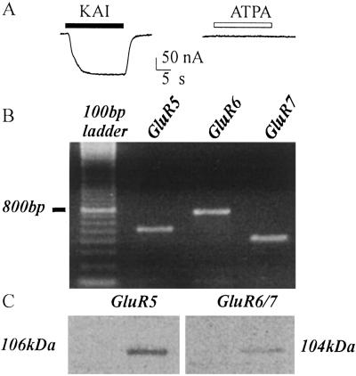
Fig 5.
Absence of functional GluR5 in TLE mRNA-injected oocytes. (A) Current response to KAI (50 μM) and lack of response to ATPA (200 μM) in the same oocyte. (B) RT-PCR analysis of GluR5, GluR6, and GluR7 subunits in TLE mRNA. (C) Western blot analysis of the expression of GluR5, GluR6/7 subunits in TLE tissue. (B and C) Same patient as in A. Note the presence of GluR5. Results are representative of all experiments from patients 1–9 of Table 1.

