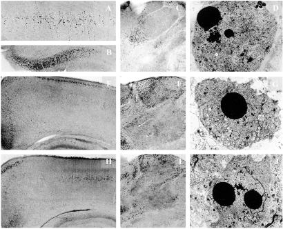
Fig 1.
Light microscopic overviews of silver-stained transverse sections and electron micrographs depicting neurodegenerative changes in the brains of P8 rats after treatment with phenytoin (A–D), diazepam (E–G), or valproate (H–J). (A–C) Layer IV of the parietal cortex (A, ×40), the subiculum (B, ×40), and the thalamus (C, ×25) of P8 rats treated 24 h previously with phenytoin (50 mg/kg). (E and F) Low-magnification (×25) light microscopic overviews of silver-stained sections from the parietal and cingulate cortices (E) and the thalamus (F) of P8 rats treated 24 h previously with diazepam (30 mg/kg). (H and I) Low-magnification (×25) light microscopic overviews of silver-stained sections from the parietal and cingulate cortices (H) and the thalamus (I) of P8 rats treated 24 h previously with valproate (200 mg/kg). Degenerating neurons (small dark dots) are sparsely present after treatment with saline in those same brain regions but abundantly present after treatment with phenytoin, diazepam, or valproate. (D, G, and J) Electron micrographs (×1,800) illustrating late stages of apoptotic neurodegeneration within the thalamus 24 h after administration of phenytoin (D), diazepam (G), or valproate (J) to rats on P7.

