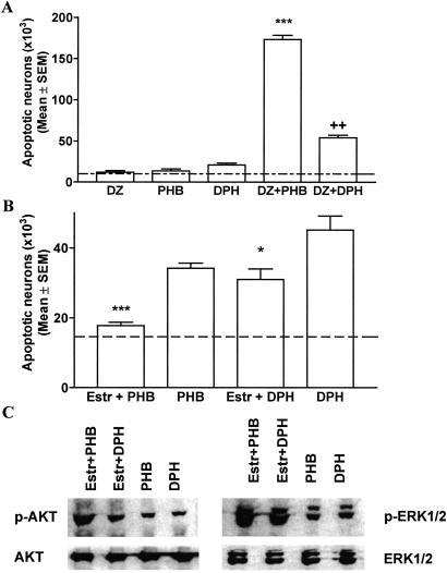
Fig 3.
(A) Combinations of AEDs elicit neurotoxic effects. Diazepam (DZ, 5 mg/kg), phenobarbital (PHB, 20 mg/kg), and phenytoin (DPH, 20 mg/kg) were administered alone or in combination to P7 rats. Severity of degeneration was established as described in Materials and Methods. Histographic values represent cumulative scores for apoptotic damage (means ± SEM, n = 6 per group) in the forebrains of treated rats on P8. Dotted line represents the mean apoptotic score in saline-treated rats. Whereas these doses of AEDs alone elicited no or minimal neurotoxic response, their combination resulted in severe apoptotic neurodegeneration (***, P < 0.001 compared with DZ- and PHB-treated rats; ††, P < 0.01 compared with DPH-treated rats, Student′s t test). (B) β-Estradiol (Estr) ameliorates apoptotic response to phenobarbital (PHB) and phenytoin in P7 rats. Saline or β-estradiol (300 μg/kg) were administered s.c. to P6 rats in three injections 8 h apart. Pups received i.p. injection of phenobarbital (50 mg/kg) or phenytoin (30 mg/kg) on P7 and the brains were analyzed on P8. Histographic values represent cumulative scores for brain damage (means ± SEM, n = 6 per group) in the forebrains of treated rats on P8. The scores illustrate that there are fewer apoptotic neurons in the brains of β-estradiol-treated rats compared with saline-treated rats (***, P < 0.001 for phenobarbital; *, P < 0.05 for phenytoin, Student′s t test). (C) β-Estradiol increases protein levels for p-ERK1/2 and p-AKT but does not alter total protein levels in the thalamus of rats treated with phenobarbital (50 mg/kg) or phenytoin (50 mg/kg). Immunoblotting was performed with antiphospho-ERK1/2, antiphospho-AKT, ERK1/2 (phosphorylation state independent), or AKT (phosphorylation state independent) antibodies. Blots are representative of a series of four blots for each antibody and each treatment condition.

