Fig. 2.
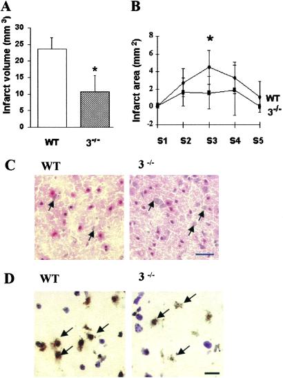
Caspase-3−/− mice were resistant to ischemia induced by 2 h of distal MCAO and 48 h of reperfusion. (A) Infarct volume in caspase-3−/− mice (n = 7) was significantly smaller than in WT littermates (n = 9; P <0.05). (B) Infarct area was decreased in caspase-3−/− mice in section 3 (S3; P < 0.05). (C) Cortical injury was less severe in caspase-3−/− mice as compared with WT littermates with more normal appearing neurons (arrows) within the lesion (hematoxylin/eosin stain). (D) Fewer TUNEL-positive cells were present in ischemic caspase-3−/− cortex than in WT littermates. Many nuclei of TUNEL-positive cells exhibited condensed or irregular nuclear clumping (arrows; counterstained with cresyl violet). (Bars = 20 μm.)

