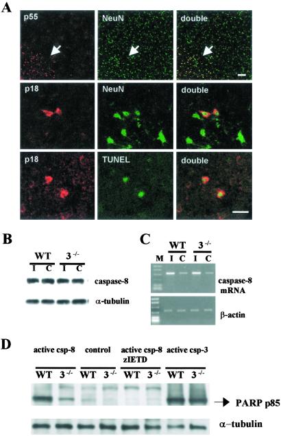
Fig. 6.
Caspase-8 expression in frontal cortex of caspase-3−/− mice after 2 h of MCAO and 24 h of reperfusion. (A Upper) Procaspase-8 (p55; red) was constitutively expressed in NeuN-positive cells (green) and was enhanced in the outer margin of the ischemic territory (arrow). Caspase-8 p18 (Middle; red) was detected within the cytoplasm of neurons (green) and colocalized with TUNEL-positive cells (green; Lower). No apparent difference occurred in the number of caspase-8 p18 cells in caspase-3−/− and WT littermate mice after ischemia (not shown). Striatal level. (Bar = 20 μm.) (B) Caspase-8 p55 was detected in ischemic (I) and contralateral (C) cortex without apparent difference (by densitometry, not shown). (C) Agarose gel showing caspase-8 mRNA up-regulated in ischemic brain (I) (Upper). No differences between strains were determined (by densitometry, not shown). β-Actin was used as a housekeeping gene (Lower). (D) Recombinant active caspase-8 (0.1 unit/μl) cleaved endogenous PARP to the 85-kDa fragment in brain homogenates of both strains, and the effect was blocked by zIETD.fmk (200 μM). Recombinant active caspase-3 (0.1 unit/μl) was used for positive control.

