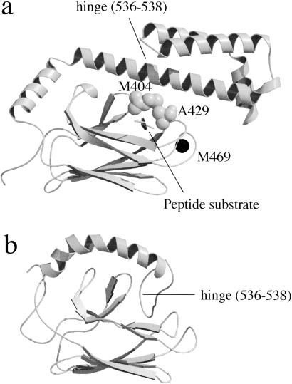
Fig 1.
3D structure of the substrate binding domain of DnaK. The secondary structure representation was drawn with molscript (33) and RASTER 3D (34). (a) DnaK389-607 complexed with a model peptide substrate solved by x-ray crystallography (3) with a space-filling model of Met-404 and Ala-429 and the position of the mutation M469C that was labeled with IAEDANS. (b) DnaK386-561 solved by NMR (22).

