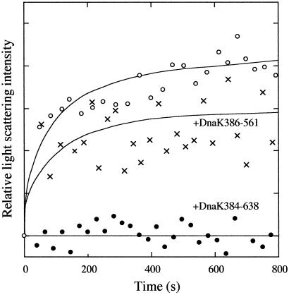
Fig 4.
Aggregation of β-galactosidase in the absence (○) and presence of 10 μM DnaK384-638 (•) or 10 μM DnaK386-561 (x). β-Galactosidase denatured with 8 M urea was diluted into refolding buffer at 10°C. The final concentrations of β-galactosidase and urea were 8.6 μM (monomer concentration) and 0.73 M, respectively. Relative light scattering at 488 nm was measured at a fixed angle of 90°.

