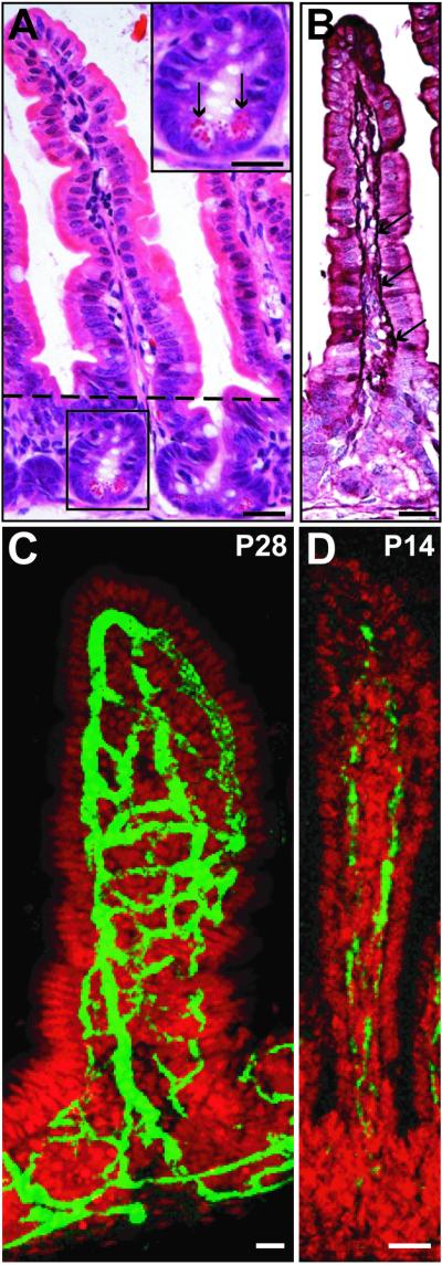
Fig 1.
Villus capillary networks in conventionally raised mice. (A–C) Sections prepared from the junction between the middle and distal thirds of the small intestine of a normal P28 mouse. (A) Hematoxylin/eosin (H&E)-stained section of a crypt–villus unit. The villus is located above, and the crypt below the dashed line. (Inset) A higher power view of the crypt showing Paneth cells (arrows). (B) Section from the same animal as in A, but stained with H&E and antibodies to the endothelial marker, von Willebrand's factor (purple; arrows). (C) Single confocal microscopic scan of a 120-μm-thick cryosection. The capillary network is stained with FITC-tagged high-molecular-weight dextran (green), and epithelial nuclei with Syto61 (red). To view a three-dimensional rotating image of a compiled set of serial scans of this type, see Movies 1 and 2, which are published as supporting information on the PNAS web site, www.pnas.org, or go to http://gordonlab.wustl.edu/vasculature. (D) Confocal view of FITC-dextran-labeled vessels in a villus positioned at the junction of the middle and distal thirds of the small intestine of a normal, conventionally raised P14 mouse. (Bars, 25 μm.)

