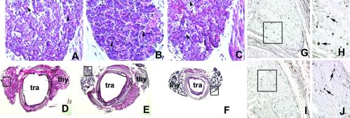
Fig 1.
Hematoxylin/eosin stain of wild-type and mutant mouse thyroid gland and BrdUrd incorporation in developing mouse thyroid gland. (A–C) Sagittal sections of E17 wild-type (A), tshrhyt/tshrhyt (B), and pit1dw/pit1dw (C) mouse embryos. The black arrowheads in A–C point to small follicles. These data were representative of at least two independent experiments performed on embryos coming from different littermates. As controls, wild-type C57BL/6 embryos were used. (D–F) Transversal sections of thyroid gland and trachea of wild-type (D), tshrhyt/tshrhyt (E), and pit1dw/pit1dw (F) of 2-month-old mice. thy, thyroid; tra, trachea. These data were representative of three independent experiments (two homozygous males and one homozygous female for both tshrhyt and pitdw strains). The mice were generated in different littermates. As controls, wild-type C57BL/6 mice were used. (G–J) Sagittal sections of E16.5 wild-type (G and H) and tshrhyt/tshrhyt (I and J) embryos. H and J show higher magnification of the boxed areas in G and H, respectively. These data are representative of three independent experiments. Embryos from untreated pregnant mice at the same days of gestation were used as a negative control. Cells that incorporated BrdUrd are visible as black dots (arrows).

