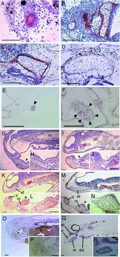
Fig 3.
Type II Collagen protein and mRNA expression in the explants and general embryos. The serial sections of the histologically analyzed explants and the sections of general embryos were immunostained with anti-Col2 antibody which was visualized with AEC and counterstained with hematoxylin. The sections of explants and general embryos were hybridized with DIG-labeled Col2 RNA probe, and the localization was visualized with NBT/BCIP. (A) Immunolocalization of Col2 in the serial section of Fig. 2B, the 4 day-cultured explant. (B) Immunolocalization of Col2 in the serial section of Fig. 2C, 7 day-cultured explant. (C) Immunolocalization of Col2 in the serial section of Fig. 2D, 10 day-cultured explant. (D) Immunolocalization of Col2 in the serial section of Fig. 2F, 14 day-cultured explant. (E) Col2 mRNA expression in the section of 4 day-cultured explant. (F) Col2 mRNA expression in the section of 7 day-cultured explant. (G) Immunolocalization of Col2 in the section of Stage 35 embryo. (H) Higher magnification of the arrowhead indicating area in G. (I) Immunolocalization of Col2 in the section of Stage 40 embryo. (J) Higher magnification of the arrowhead indicating area in I. (K) Immunolocalization of Col2 in the section of Stage 42 embryo. (L) Higher magnification of the arrowhead indicating area in K. (M) Immunolocalization of Col2 in the section of Stage 44 embryo. (N) Higher magnification of the arrowhead indicating area in M. (O) Col2 mRNA expression in the section of Stage 35 embryo. (P) Higher magnification of indicating area in O. (Q) Col2 mRNA expression in the section of Stage 44 embryo. (R) Higher magnification of indicating area in Q. et, ethmoid trabecular cartilage; ir, infrarostral cartilage; m, Meckel's cartilage; nt, notochord; pq, palatoquadrate cartilage. (Bars = 50 μm.)

