Fig 3.
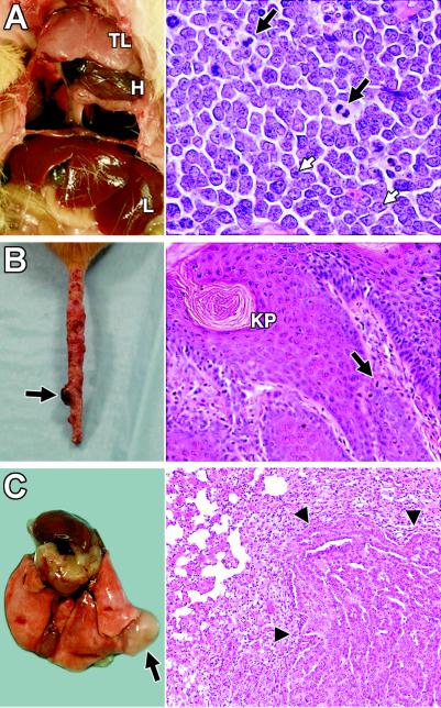
Tumors in Pold1D400A/D400A mice. (A) Mediastinal thymic lymphoma (TL). H, heart; L, liver. Lungs are collapsed and not visible. (Right) Hematoxylin and eosin (H&E)-stained section of thymic lymphoma (original magnification, ×400). White arrows, nucleoli; black arrows, mitotic figures. (B) Tail with marked irregularities throughout its length and a focal skin tumor (arrow). (Right) Section of skin tumor stained with H&E (original magnification, ×200). KP, keratin pearl; arrow, mitotic figure. (C) Lung adenocarcinoma (arrow). (Right) H&E-stained section showing the tumor (arrowheads) compressing the normal lung parenchyma (original magnification, ×100).

