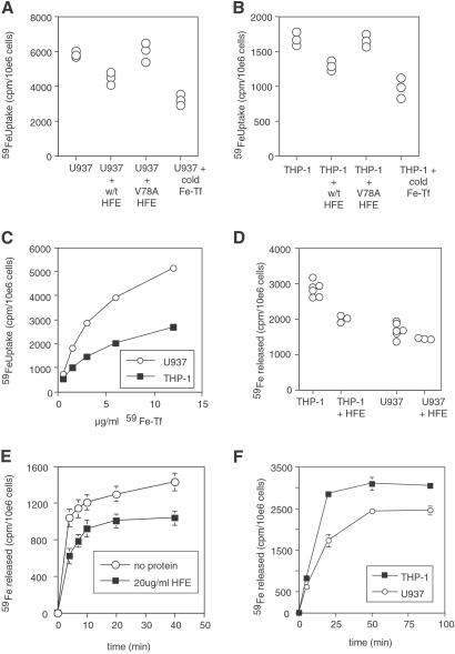
Fig 2.
Effect of HFE on iron uptake and release. (A and B) WT HFE blocks iron uptake into THP-1 and U937 cells. Log-phase U937 (A) and THP-1 (B) cells were grown with 27 nM 59Fe-Tf for 6 h in the presence of 270 nM cold human holo-Tf, 300 nM soluble HFE, or 300 nM soluble V78A HFE as indicated, and then cellular 59Fe was measured. Each point represents the uptake into one aliquot of 106 cells. (C) Comparison of iron uptake by U937 and THP-1 cells. Log phase U937 and THP-1 cells at 106 cells per ml were incubated with given doses (10 μg/ml = 133 nM) of 59Fe-Tf for 4 h, and then cellular 59Fe was measured. (D) Effect of HFE on iron release by U937 and THP-1 cells. Cells were grown in 53 nM 59Fe-Tf for 2 days, then washed and allowed to export iron into media for 45 min in the presence of 300 nM HFE protein as shown. Each point represents the release of one aliquot of 106 cells. (E) Time course of iron release from THP-1 cells in the presence or absence of HFE. THP-1 cells were loaded with iron, as in C, and allowed to export iron for the times indicated with or without addition of soluble HFE (300 nM) to the medium. Error bars indicate the range of values in the triplicate samples for each condition. (F) Time course of iron release from U937 and THP-1 cells. Cells were grown in 53 nM 59Fe-Tf for 2 days, then washed and allowed to export iron into media for times shown. Error bars indicate the range of values in the triplicate samples for each condition.

