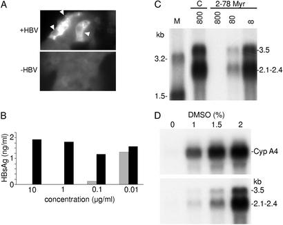
Fig 4.
Specificity of the HBV infection. (A) Immunostaining of infected HepaRG cells for HBV core antigen. Cells were stained with an anticore antibody by immunofluorescence. Positive cells are indicated by a white arrow. (B) In vitro neutralization assay. Viral particles were incubated with serial dilutions of a monoclonal anti-HBs antibody (S 39–10;  ) or an unrelated monoclonal antibody (▪) and evaluated for their infectivity on HepaRG cells. Infectivity was evaluated by measuring the HBsAg secretion in the supernatant of infected cells at day 10 pi. (C) Peptide competition of HBV infection. HepaRG cells were preincubated with different concentrations (nM) of an HBV large protein-derived peptide (AA 2–78) bearing a myristic acid (myr) on gly 2 or, as a control, with a corresponding myristoylated duck hepatitis B virus-derived peptide (AA 2–41; C). The inoculum was then added for 20 h, without removing the peptide. Infectivity was evaluated by analyzing intracellular viral RNA at 10 days pi. Half of the RNA extracted from 106 infected cells was analyzed on a 1.5% agarose gel. Lane M corresponds to 3.5 pg of denatured 3.2- and 1.5-kb HBV DNA restriction fragments. Viral RNA sizes are indicated on the right. Autoradiographic exposure time was 48 h. (D) Efficiency of HBV infection and HepaRG cell differentiation. Northern blot analysis of CYP 3A4 (Upper) and HBV (Lower) RNAs after infection of HepaRG cells cultivated in presence of increasing concentrations of DMSO. RNA was extracted 10 days pi. Viral RNA sizes are indicated on the right.

