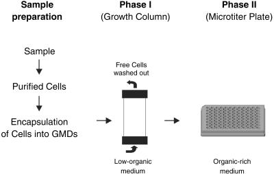
Fig 1.
Model of the experimental setup. Cells captured from environmental samples were encapsulated into GMDs and incubated in growth columns (phase I). GMDs containing microcolonies were detected and separated by flow cytometry into 96-well microtiter plates containing a rich organic medium (phase II).

