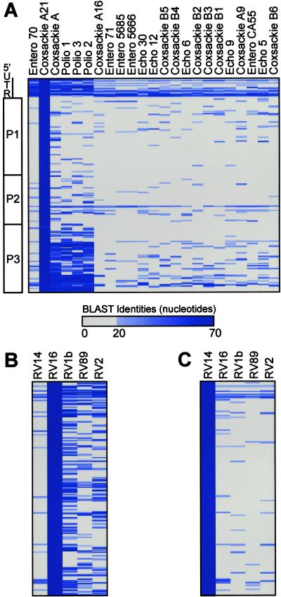
Fig 1.
(A) Graphical depiction of homology between coxsackie A21 and other enteroviruses. Picornavirus genomic organization is shown (Left). Seventy-nucleotide segments from the coxsackie A21 genome are ordered sequentially downward from the 5′ end of the genome. The number of nucleotides of identity between each 70-nt segment (rows) and each virus (columns) in the genus enterovirus is reflected by the intensity of the blue bar. Identities of <20 nt were plotted as gray. (B) Homology between RV16 and other RVs. (C) Homology between RV14 and other RVs.

