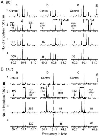
Fig 2.
Changes in the direction of BF shifts of a collicular and a cortical DSCF neuron of the mustached bat. BMI applied to cortical DSCF neurons changes centrifugal BF shifts (a) evoked by ES of cortical DSCF neurons into centripetal BF shifts (b and c). The arrays of PST histograms display frequency–response curves of a collicular (A) and a cortical (B) DSCF neuron. The vertical and horizontal arrows respectively indicate the BFs of cortical DSCF neurons receiving ES and/or BMI and centrifugal or centripetal BF shifts of the recorded neurons. 1–4: arrays of PST histograms recorded before (control) and after ES and/or BMI applications. The amplitude of tone bursts was set at 10 dB above the minimum threshold of a given neuron. ES, 0.2-ms 100-nA electric pulses delivered at a rate of 5/s for 7.0 min; BMI, 1.0 nl of 5 mM BMI.

