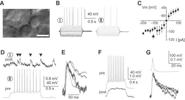
Fig 3.
Spiny projection neurons in acute striatal slices are connected through a monosynaptic, fast synaptic connection. (A) Acute striatal slice with contiguous medium-sized, spherical cell bodies during dual whole-cell patch recording. (Bar = 10 μm.) (B) Responses of spiny projection neurons to current injections (VRest I = −62 mV and VRest II = −68 mV). (C) I–V relationship for spiny projection neurons from acute slices (n = 89). Note the strong anomalous rectification below −90 mV (filled arrow). (D) Synaptic connection between spiny projection neurons in acute striatal slices (same neurons as in B). Note the high failure rate. (E) Overplot of average responses (failures excluded) for all five connected pairs normalized by amplitude. (F) Electrical synapse in a pair of spiny projection neurons (acute slice; VRest pre = −62 mV and VRest post = −74 mV). (G) Typical spikelet in electrically coupled spiny projection neurons at three different membrane potentials (−70, −60, and −50 mV). pre, presynaptic; post, postsynaptic.

