
FIGURE 2.
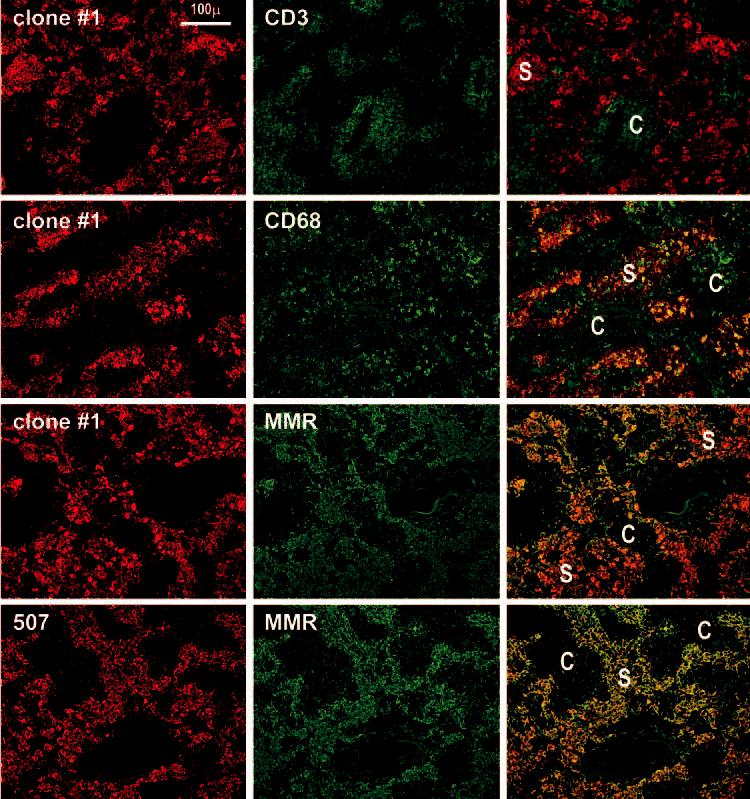
DC-SIGN staining is restricted to macrophages in the medullary sinuses (S) of lymph nodes. Two-color staining is shown, with red being the anti-DC-SIGN reagent, and green the other markers. First row, Clone 1 anti-DC-SIGN stains macrophages in the sinuses of the medulla and anti-CD3 stains T cells in the medullary cords (C). Second row, Clone 1 and anti-CD68 colabel medullary macrophages. Third row, Clone 1 and anti-macrophage mannose receptor (MMR) colabel macrophages in the lymph node medulla. Fourth row, Double staining using a commercial anti-DC-SIGN 507 and anti-mannose receptor mAbs.