
FIGURE 4.
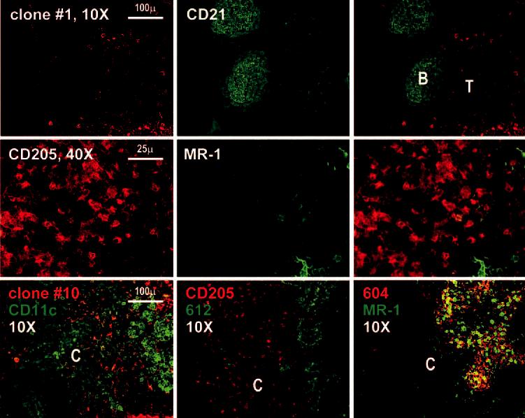
Immunostaining of the lymph node cortex with anti-DC-SIGN mAbs. First row, The lymph node cortex is shown at ×10 after staining with anti-CD21 (green) to define the B cell follicles (B) and the unstained T cell region (T). Second row, Images at higher magnification within the T cell region reveal that most DCs defined by staining with anti-CD205 are DC-SIGN negative. Third row, The left panel is a double staining with our clone 10 (specific for DC-SIGN and L-SIGN) and CD11c; the middle panel is a double staining with clone 612 (specific for DC SIGN and L-SIGN) and DEC205; the right panel shows a double staining using anti-L-SIGN specific mAb (604) with MR-1.