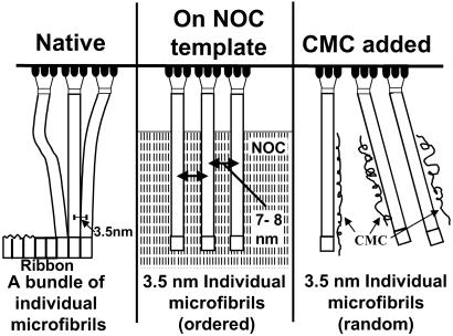
Fig 5.
Schematic diagram comparing three patterns of ribbon assembly. The enzyme sites from the bacterial surface are shown at the top, with aggregating glucan chains to form three microfibrils. Native condition (Left) occurs when the cell is freely suspended in the growth medium and produces a tight ribbon of microfibrils that are closely associated with each other. Ribbons produced in association with the NOC template track (Center) tightly adhere to the substrate and are not as tightly aggregated as in the native state. With the addition of CMC, individual microfibrils are immediately coated and prevented from aggregation (Right).

