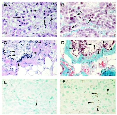
Fig 7.
Histological and immunocytochemical analysis of tumors from nude mice. (A) Untreated control UISO-Mel-2 flank tumor 3 weeks after inoculation in nude mice. Abundance of viable malignant cells as shown by arrows. [Hematoxylin/eosin (H&E) stain, ×40.] (B) Untreated control UISO-Mel-2 tumor 3 weeks after inoculation. Masson's trichrome selectively stains fibrous tissues blue; a normal distribution of blue-stained fibrous tissues with similar numbers of malignant cells as in A is seen (shown by arrows). (×40.) (C) Azurin-treated UISO-Mel-2 tumor after 3 weeks of treatment. Apparent is a paucity of viable malignant cells but with many nonviable (apoptotic) cells (arrowhead) with areas of regression and fibrosis, stained deep purple with H&E stain. (×40.) (D) Same azurin-treated UISO-Mel-2 tumor after 3 weeks of treatment. Stained with Masson's trichrome stain. Many nonviable (apoptotic) cells (shown by arrowhead) are seen. The extensive blue areas of fibrosis are pronounced. Similar to C but in contrast to B, very few viable melanoma cells are observed. (×40.) (E) Untreated control 3 weeks after inoculation in nude mice. TUNEL stain shows scanty apoptotic cells. In this section, only one is easily recognizable (shown by arrowhead). (×40.) (F) Azurin-treated tumor cells, 3 weeks later. TUNEL stain shows a multitude of apoptotic cells (shown by arrowheads). (×40.)

