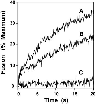
Fig 4.
Contact time between the GAPDH isoform-catalyzing membrane fusion and tubulin as a determinant of fusion velocity. Physiologically modeled SUVs were prepared and loaded into one chamber of a stopped-flow apparatus as described in Experimental Procedures. The other chamber was loaded with the membrane fusion-catalyzing protein from the GTP-agarose column diluted to a concentration of 1 μg/ml (A). Purified tubulin (5 μg/ml) was preincubated with SUVs for 30 s at 37°C before mixing in the stopped-flow apparatus with the GAPDH-containing solution (1 μg/ml) (B). Tubulin (5 μg/ml) and the GAPDH isoform-catalyzing membrane fusion (1 μg/ml) were placed in one chamber, and vesicles were placed in the other chamber; fusion was initiated by rapid mixing of chambers and quantified by the increase in R18 fluorescence as described in Experimental Procedures (C).

